
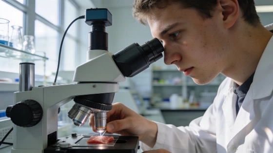
Picture a doctor able to look inside your body and pick out the cells that have stopped dividing and started causing age-related problems. MIT scientists have just taken a big step toward that goal. They have built a noninvasive way to find senescent cells—often called "zombie cells"—that pile up as we age and drive diseases like cancer and diabetes.
External summaries of the study report that the RamanOmics barcode classifier achieved an estimated accuracy of 94.7% in distinguishing senescent cells from healthy ones.
Until now, finding these cells meant looking for markers like the proteins p16 and p21, but those tests destroy the cells being studied. RamanOmics avoids that. It shines near-infrared or visible light on living cells to catch their chemical signals, then adds spatial RNA sequencing to map gene activity. This builds a detailed profile that can tell senescent cells apart from healthy ones in real time.
In their study in Nature Aging, the MIT team used RamanOmics on skin and lung tissue from young (2-month-old) and old (26-month-old) mice. The results showed big changes in older cells, especially a jump in lipid production and buildup. In skin, senescent cells had changes in muscle contraction and collagen remodeling pathways. In old lung tissue, there was more immune activity and inflammation. These patterns show how aging works differently in each tissue. As Scientific Frontline coverage notes, the research is still preclinical and has only been tested in mice, not humans.
The concept of senescent cells as accumulating 'zombie cells' has long been linked to age-related diseases. The new MIT approach is significant because it moves beyond destructive markers like p16/p21, instead leveraging Raman microscopy and single-cell gene expression profiling to noninvasively map senescence.
What makes RamanOmics different is its barcode—a mix of Raman spectral peaks and gene signatures—that marks senescent cells. "Combining the most important Raman features with the most important gene signatures, we were able to create a barcode that can help us to identify senescent cells in a more unbiased way," said Salvatore Sorrentino, PhD, co-first author. This barcode could let doctors quickly and safely check tissue aging or test senolytic treatments in living patients.
The current study used mouse tissue, but the team is already working to adapt the method for human samples. They are also building a faster Raman imaging system, hoping to cut the current 30-hour scan time for a one-square-millimeter sample to something much quicker for clinics.
Cellular senescence is a double-edged sword. It leads to tissue breakdown and chronic disease as we age, but it also helps in development and wound healing. As Peter So said, "Senescence is not just a pathological condition." The NIH’s broad approach aims to map senescence in both healthy and diseased states.
RamanOmics is more than a research tool. It could change how we diagnose, track, and treat aging. By making these hidden cells visible, the method could push the field past destructive tests and toward real-time checks in living tissue. The next step is to move from mouse models to human medicine, but the groundwork is set for a new chapter in longevity science.