
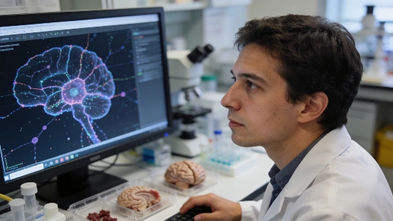
Inside a Madison lab, researchers have started charting Alzheimer’s disease one cell at a time. Their new AI-powered tools sift through millions of brain cells, exposing which genes and cell types tie directly to symptoms and cognitive resilience in each patient.
Alzheimer’s rarely follows a script. Symptoms and progression jump from person to person, leaving traditional research methods scrambling to separate the drivers from the noise. Daifeng Wang’s team at UW–Madison turned to graph neural networks, linking clinical symptoms to the underlying biology at single-cell scale. UCI MIND’s independent review places this work within the PsychAD Consortium and confirms the use of the largest gene expression map of the human prefrontal cortex to date—over 6 million cells from nearly 1,500 donors.
The project used single-nucleus RNA sequencing, a technique that enables researchers to assess gene activity at the level of individual cells, providing unprecedented resolution for studying brain disorders.
PASCode and iBrainMap in action
The team built PASCode and iBrainMap using data from 6.3 million brain cells across nearly 1,500 autopsy donors. PASCode scores each cell for its link to Alzheimer’s symptoms, including depression and cognitive decline. About 1.5 million cells landed in the Alzheimer’s-associated group, letting researchers zero in on the cell populations and gene shifts that matter most for disease progression and resilience. The UCI MIND project overview notes the prefrontal cortex was chosen for its central role in memory and decision-making, making it a prime target for studying Alzheimer’s effects and resilience.
iBrainMap digs deeper, building a gene roadmap for each donor. It tracks how DNA mutations disrupt gene networks in individual brains, flagging cell-type specific genes that may shape Alzheimer’s course and neuropsychiatric symptoms like weight loss or insomnia. This approach boosted the accuracy of distinguishing healthy from Alzheimer’s-affected brains by 21% over older methods. UCI MIND’s review confirms the project’s scale and methods but does not independently verify the 21% improvement or clinical effectiveness of PASCode and iBrainMap.
Individualized gene signatures and new subtypes
Instead of sorting patients into broad groups, the researchers mapped out symptom-specific gene lists, cell types, and gene networks for each donor. This level of detail uncovered new Alzheimer’s subtypes, hinting that some patients share unique molecular and cellular features that could be targeted with custom therapies.
The team published all findings in a public atlas, giving other scientists a way to explore disease-linked cell types and gene networks in their own data. The project sits within the PsychAD Consortium, a National Institutes of Health-backed effort led by Wang and Panos Roussos at Mount Sinai Medical School, focused on finding biomarkers and therapeutic targets for Alzheimer’s and related neuropsychiatric symptoms.
The brain cell map resource was published on October 6, 2026, and updated on October 7, 2026. UCI MIND emphasizes that this atlas is intended as a research tool and is not approved for clinical diagnostic use. No regulatory decisions or clinical guidelines currently authorize the use of PASCode or iBrainMap for patient diagnosis, prognosis, or treatment selection.
Operational impact for research and therapy
Mapping Alzheimer’s at single-cell resolution with AI tools gives researchers a sharper lens for diagnosis and the chance to pursue personalized interventions. The discovery of new subtypes and patient-specific gene targets signals a move away from blanket approaches, pushing the field toward therapies that fit each patient’s biology.
This technical leap echoes earlier innovations that expanded the toolkit for decoding complex biology. The Alzheimer’s atlas now stands as a resource for labs worldwide. So far, the evidence is computational and bioinformatic, not clinical. The atlas remains a research tool, with no regulatory approval for diagnosis or treatment.