
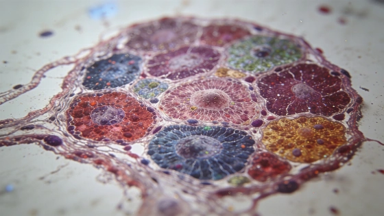
Inside a lymph node, an immune cell’s location may affect how it survives and responds to threats. That question drives Cells and the Suburbs, the overall winner of WEHI’s 2026 Art of Science exhibition. The work is a colour-segmented map of thousands of cells in a mouse lymph node.
Dr Brigette Duckworth and Dr Chin Wee Tan created the image. It shows distinct cellular neighborhoods occupied by memory T cells, which remain in the body after infection and provide long-term disease protection. Studying the environments around those cells could help researchers identify targets for vaccine design and cancer immunotherapy.
In cancer immunology, spatial profiling can help researchers study immune-cell infiltration, immune exclusion and treatment resistance in the tumor microenvironment, and explore potential biomarkers and treatment combinations.
Reading cells in their neighborhoods
The image uses spatial transcriptomics to connect genetic information from thousands of cells with their locations in tissue. In general, the method links gene activity to tissue coordinates, letting researchers examine cellular interactions and local environments alongside the cell types present. A spatial immune-mapping study describes the approach.
Duckworth says location can shape a cell’s survival and function. In a lymph node, an immune cell’s position can influence how effectively it responds to a threat such as a viral infection. Mapping the whole tissue can reveal patterns that a cell-by-cell view might miss.
Lymph nodes are specialized immune environments where antigen-presenting cells can take up vaccine material and activate T cells. Delivering vaccine components to draining lymph nodes is being explored as a way to support antigen presentation and immune responses; this is research context, not evidence of a clinical benefit from the map described here.
The research does not establish a new vaccine or show improved treatment outcomes. Spatial mapping can help researchers chart immune-cell distribution and investigate potential biomarkers. But a biomarker-focused review in immunotherapy research stresses that these methods alone cannot show that a vaccine works better or that patients have better outcomes. The possible applications of this work remain a research prospect.
Making research visible
Now in its 29th year, WEHI’s online exhibition features 12 finalists. Their work spans infectious diseases and cancer. Other projects cover inflammation, while further entries explore neurodegenerative diseases and structural biology. The works use scientific experiments and advanced imaging to make research processes visible. Data visualisation is another tool in the exhibition. A Blavatnik Awards report follows scientists working in gene editing and chemistry. It also covers bioelectronics; WEHI’s exhibition instead puts a research image at the center of the story.
The image makes a research question tangible: how does a cell’s neighborhood affect what it can do? That offers a clear way to show the public how biologists study immunity, where spatial context can matter as much as cell identity. External results did not independently corroborate the specific artwork, artist or award details. A separate WEHI exhibition page covered a different work, so those details remain as reported in the exhibition account.